



A Clear Guide to DEXA Scans and Bone Health
News
/
November 4, 2026
What Does a DEXA Scan Show, and How Do You Prepare for One?
A Clear Guide to DEXA Scans and Bone Health
Body contouring has revolutionized the way we think about self-care and aesthetic transformations. It’s more than just a cosmetic procedure—it’s a path to enhanced confidence and self-expression.
What a DEXA Scan Measures
A DEXA scan, also called a DXA scan, is a common test for measuring bone mineral density. The name stands for dual-energy X-ray absorptiometry. It uses low-dose X-rays to measure calcium and other minerals in bones, usually at the hip and lower spine.
Bones with more mineral content are generally denser and stronger, which can help clinicians assess bone health and estimate fracture risk. A DEXA scan can support evaluation for conditions such as osteopenia and osteoporosis, and help monitor changes over time. It is not the same as a bone scan, which uses an injection to investigate other problems. The National Institute of Arthritis and Musculoskeletal and Skin Diseases explains how bone mineral density tests work.
The result is one part of a clinical evaluation, not a complete explanation of bone health. A scan can show low density, but it cannot identify its cause or determine fracture risk on its own. Clinicians interpret the findings alongside a person’s health history and other risk factors. Mayo Clinic’s overview of bone density testing describes its uses and limits.
Scan Sites and Related Tests

A DEXA result helps clinicians assess bone health, rather than simply produce an image. It can reveal low bone density, support evaluation for osteopenia or osteoporosis, estimate fracture risk, and show changes in bone density over time or in response to treatment (NIAMS guidance on bone mineral density tests).
Most central scans measure the hip and lower spine, common sites of fractures associated with osteoporosis. Depending on the clinical question, the forearm may also be examined. At a Manhattan practice such as thomasloebmd.com, care is planned around each patient’s individual needs; decisions about bone testing, however, belong with the patient’s medical clinician.
Central and peripheral scans
A central DEXA scan measures the hip or spine. Peripheral tests assess areas such as the finger, wrist, heel, or forearm. These smaller-area tests can help with screening, but they are generally less detailed or accurate than central scans. If a peripheral result suggests low density, a clinician may recommend a hip or spine scan for further assessment (Mayo Clinic’s overview of DEXA testing).
DEXA is also different from a whole-body bone scan. DEXA measures bone density without an injection. A bone scan involves an injected substance and may be used to investigate concerns such as infection, cancer, or fractures. The two tests answer different clinical questions, so the appropriate examination depends on what a clinician needs to assess.
Who May Need Bone Density Testing

Whether to have a bone density test depends on age, medical history, medications, and fracture risk. A clinician can weigh these factors together rather than relying on one risk factor alone. The National Institute of Arthritis and Musculoskeletal and Skin Diseases notes that screening recommendations differ by group, including for men.
Commonly cited groups for testing include women age 65 or older, postmenopausal women younger than 65 who have increased fracture risk, men age 70 or older, and people over 50 who have broken a bone. The U.S. Preventive Services Task Force recommends screening for older women and younger postmenopausal women at increased risk, but says evidence is insufficient to recommend for or against routine screening in men.
A clinician may also consider testing after substantial height loss, a fracture from a minor fall or other low-force event, long-term use of steroid medicines such as prednisone, or a drop in hormone levels after menopause or certain cancer treatments. These circumstances can signal a need to assess bone density, but do not determine a diagnosis on their own.
Other factors that may raise concern include low body weight, a family history of osteoporosis, smoking, limited physical activity, and inadequate calcium or vitamin D. Some health conditions and medications can also affect bone strength. The Mayo Clinic’s guidance on bone density testing describes several circumstances clinicians may consider, while emphasizing that the scan is only one part of evaluating bone health.
What to Expect During the Exam
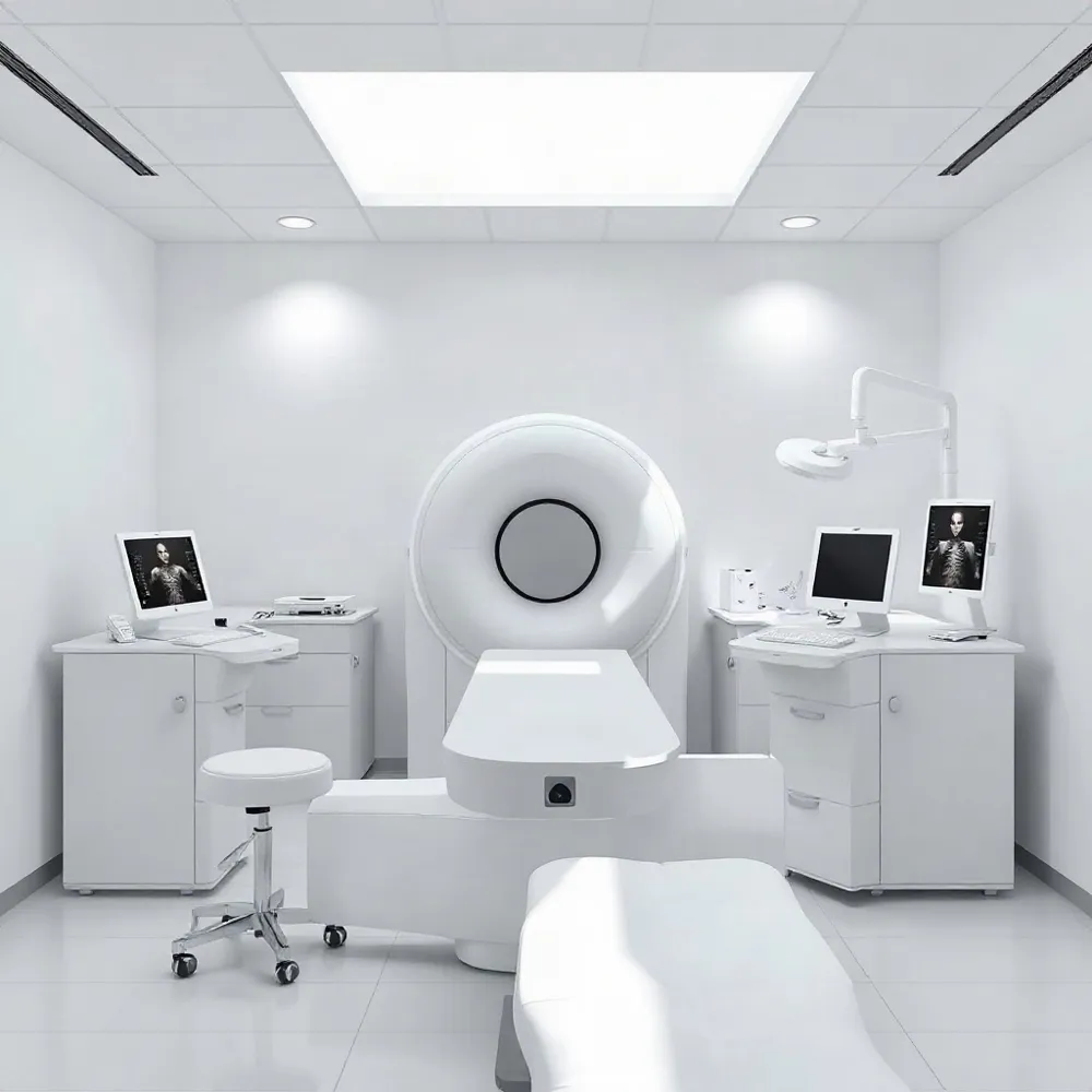
A DEXA scan is usually an outpatient exam. You lie on a padded X-ray table while a technician positions you and moves a scanning arm over the area being checked. Foam supports may be used to help position your legs for spine or hip images. The setup is open, not an enclosed tube, as described in this guide to DEXA scans.
The scan is generally painless and does not involve needles or injections. You will need to remain still while images are taken so movement does not affect them. The technician may give specific directions about your position or when to hold still, and procedures can vary somewhat between imaging centers.
A typical examination takes about 10 to 30 minutes, depending on which areas are scanned. The X-ray exposure is very low, but tell your healthcare professional if you are pregnant or might be pregnant. DEXA testing is generally avoided during pregnancy when possible, so the care team can discuss whether and when to proceed.
Most people can return to their usual activities after the scan. Your clinician can explain when and how you will receive the results.
How to Prepare for a DEXA Scan
Preparation is usually minimal. Most people can eat, drink, and take their regular medicines as usual unless their clinician or imaging center gives different instructions. Follow the directions provided for your appointment, since requirements can vary.
If you take calcium supplements or other over-the-counter vitamins, ask when to pause them. Guidance commonly recommends avoiding calcium supplements for 24 hours before a scan, while some instructions allow a 24-to-48-hour interval. Confirm the timing with your care team rather than assuming one rule applies to every facility.
Tell the scheduler or imaging staff about recent or planned imaging tests, especially a barium examination or contrast used for a CT or nuclear medicine test. These may affect when the DEXA scan can be performed or how its results are interpreted. Mayo Clinic’s DEXA guidance advises patients to report these tests before the scan.
Choose loose, comfortable clothing without metal fasteners, such as zippers, buttons, or belts. You may need to remove jewelry and metal objects from your pockets, and the facility may provide a gown if needed. MedlinePlus guidance on bone density scans also recommends avoiding metal jewelry and clothing parts.
Do not stop or change prescribed medicines unless your clinician tells you to. If you are unsure about supplements, recent imaging, clothing, or any other preparation detail, check with the ordering clinician or imaging facility before your appointment.
Understanding Scores and Test Limits

A DEXA report commonly includes a T-score and, in some situations, a Z-score. The T-score compares bone density with that of a healthy young adult. The Z-score compares it with results for people with similar characteristics, such as age and sex. Which score is used depends partly on age and clinical context, so a clinician can explain which comparison applies to your report.
For adults assessed with a T-score, a result of -1 or higher is generally considered normal. A result below -1 and above -2.5 indicates low bone density, often called osteopenia, while -2.5 or lower falls in the osteoporosis range. These categories are commonly used to describe results, but they are not a complete assessment of an individual’s health. NIAMS explains how bone mineral density scores are interpreted.
A lower T-score is associated with greater fracture risk, but the result alone does not predict whether a fracture will occur. Nor does a scan explain why bone density is low or determine which treatment is appropriate. A clinician considers the score alongside factors such as medical history and fracture risk, and may order other tests to investigate possible causes.
Some conditions can make measurements less reliable. Arthritis, scoliosis, other spinal changes, or previous spine surgery may affect results from scans of the spine or hip. Readings can also differ between DXA machine manufacturers. When comparing scans over time, clinicians consider the equipment used and the person’s overall clinical picture, rather than treating small changes as meaningful on their own.
Coverage is another practical consideration. Medicare and some insurance plans may cover a bone density test every two years for people at risk, but eligibility and benefits vary. Check your plan before scheduling, and ask the ordering clinician whether the timing and location of the test fit your circumstances. Mayo Clinic’s guidance on bone density testing also notes that coverage depends on the individual plan.
Discuss Results with Your Clinician
A DEXA scan provides useful information about bone density, but its result is only one part of assessing bone health. Its meaning depends on factors such as age, medical history, prior fractures, and overall fracture risk. A scan can identify low density, but it cannot explain its cause or determine by itself which treatment is appropriate.
Review the report with the healthcare professional who ordered the test. Ask how the findings relate to your health and whether additional evaluation, follow-up testing, or treatment is recommended. Your clinician can interpret the results alongside other risk factors and advise whether monitoring or another scan is needed. Follow their guidance and raise questions about unclear terms or next steps.
What a DEXA Scan Measures
A DEXA scan, also called a DXA scan, is a common test for measuring bone mineral density. The name stands for dual-energy X-ray absorptiometry. It uses low-dose X-rays to measure calcium and other minerals in bones, usually at the hip and lower spine.
Bones with more mineral content are generally denser and stronger, which can help clinicians assess bone health and estimate fracture risk. A DEXA scan can support evaluation for conditions such as osteopenia and osteoporosis, and help monitor changes over time. It is not the same as a bone scan, which uses an injection to investigate other problems. The National Institute of Arthritis and Musculoskeletal and Skin Diseases explains how bone mineral density tests work.
The result is one part of a clinical evaluation, not a complete explanation of bone health. A scan can show low density, but it cannot identify its cause or determine fracture risk on its own. Clinicians interpret the findings alongside a person’s health history and other risk factors. Mayo Clinic’s overview of bone density testing describes its uses and limits.
Scan Sites and Related Tests
A DEXA result helps clinicians assess bone health, rather than simply produce an image. It can reveal low bone density, support evaluation for osteopenia or osteoporosis, estimate fracture risk, and show changes in bone density over time or in response to treatment (NIAMS guidance on bone mineral density tests).
Most central scans measure the hip and lower spine, common sites of fractures associated with osteoporosis. Depending on the clinical question, the forearm may also be examined. At a Manhattan practice such as thomasloebmd.com, care is planned around each patient’s individual needs; decisions about bone testing, however, belong with the patient’s medical clinician.
Central and peripheral scans
A central DEXA scan measures the hip or spine. Peripheral tests assess areas such as the finger, wrist, heel, or forearm. These smaller-area tests can help with screening, but they are generally less detailed or accurate than central scans. If a peripheral result suggests low density, a clinician may recommend a hip or spine scan for further assessment (Mayo Clinic’s overview of DEXA testing).
DEXA is also different from a whole-body bone scan. DEXA measures bone density without an injection. A bone scan involves an injected substance and may be used to investigate concerns such as infection, cancer, or fractures. The two tests answer different clinical questions, so the appropriate examination depends on what a clinician needs to assess.
Who May Need Bone Density Testing
Whether to have a bone density test depends on age, medical history, medications, and fracture risk. A clinician can weigh these factors together rather than relying on one risk factor alone. The National Institute of Arthritis and Musculoskeletal and Skin Diseases notes that screening recommendations differ by group, including for men.
Commonly cited groups for testing include women age 65 or older, postmenopausal women younger than 65 who have increased fracture risk, men age 70 or older, and people over 50 who have broken a bone. The U.S. Preventive Services Task Force recommends screening for older women and younger postmenopausal women at increased risk, but says evidence is insufficient to recommend for or against routine screening in men.
A clinician may also consider testing after substantial height loss, a fracture from a minor fall or other low-force event, long-term use of steroid medicines such as prednisone, or a drop in hormone levels after menopause or certain cancer treatments. These circumstances can signal a need to assess bone density, but do not determine a diagnosis on their own.
Other factors that may raise concern include low body weight, a family history of osteoporosis, smoking, limited physical activity, and inadequate calcium or vitamin D. Some health conditions and medications can also affect bone strength. The Mayo Clinic’s guidance on bone density testing describes several circumstances clinicians may consider, while emphasizing that the scan is only one part of evaluating bone health.
What to Expect During the Exam
A DEXA scan is usually an outpatient exam. You lie on a padded X-ray table while a technician positions you and moves a scanning arm over the area being checked. Foam supports may be used to help position your legs for spine or hip images. The setup is open, not an enclosed tube, as described in this guide to DEXA scans.
The scan is generally painless and does not involve needles or injections. You will need to remain still while images are taken so movement does not affect them. The technician may give specific directions about your position or when to hold still, and procedures can vary somewhat between imaging centers.
A typical examination takes about 10 to 30 minutes, depending on which areas are scanned. The X-ray exposure is very low, but tell your healthcare professional if you are pregnant or might be pregnant. DEXA testing is generally avoided during pregnancy when possible, so the care team can discuss whether and when to proceed.
Most people can return to their usual activities after the scan. Your clinician can explain when and how you will receive the results.
How to Prepare for a DEXA Scan
Preparation is usually minimal. Most people can eat, drink, and take their regular medicines as usual unless their clinician or imaging center gives different instructions. Follow the directions provided for your appointment, since requirements can vary.
If you take calcium supplements or other over-the-counter vitamins, ask when to pause them. Guidance commonly recommends avoiding calcium supplements for 24 hours before a scan, while some instructions allow a 24-to-48-hour interval. Confirm the timing with your care team rather than assuming one rule applies to every facility.
Tell the scheduler or imaging staff about recent or planned imaging tests, especially a barium examination or contrast used for a CT or nuclear medicine test. These may affect when the DEXA scan can be performed or how its results are interpreted. Mayo Clinic’s DEXA guidance advises patients to report these tests before the scan.
Choose loose, comfortable clothing without metal fasteners, such as zippers, buttons, or belts. You may need to remove jewelry and metal objects from your pockets, and the facility may provide a gown if needed. MedlinePlus guidance on bone density scans also recommends avoiding metal jewelry and clothing parts.
Do not stop or change prescribed medicines unless your clinician tells you to. If you are unsure about supplements, recent imaging, clothing, or any other preparation detail, check with the ordering clinician or imaging facility before your appointment.
Understanding Scores and Test Limits
A DEXA report commonly includes a T-score and, in some situations, a Z-score. The T-score compares bone density with that of a healthy young adult. The Z-score compares it with results for people with similar characteristics, such as age and sex. Which score is used depends partly on age and clinical context, so a clinician can explain which comparison applies to your report.
For adults assessed with a T-score, a result of -1 or higher is generally considered normal. A result below -1 and above -2.5 indicates low bone density, often called osteopenia, while -2.5 or lower falls in the osteoporosis range. These categories are commonly used to describe results, but they are not a complete assessment of an individual’s health. NIAMS explains how bone mineral density scores are interpreted.
A lower T-score is associated with greater fracture risk, but the result alone does not predict whether a fracture will occur. Nor does a scan explain why bone density is low or determine which treatment is appropriate. A clinician considers the score alongside factors such as medical history and fracture risk, and may order other tests to investigate possible causes.
Some conditions can make measurements less reliable. Arthritis, scoliosis, other spinal changes, or previous spine surgery may affect results from scans of the spine or hip. Readings can also differ between DXA machine manufacturers. When comparing scans over time, clinicians consider the equipment used and the person’s overall clinical picture, rather than treating small changes as meaningful on their own.
Coverage is another practical consideration. Medicare and some insurance plans may cover a bone density test every two years for people at risk, but eligibility and benefits vary. Check your plan before scheduling, and ask the ordering clinician whether the timing and location of the test fit your circumstances. Mayo Clinic’s guidance on bone density testing also notes that coverage depends on the individual plan.
Discuss Results with Your Clinician
A DEXA scan provides useful information about bone density, but its result is only one part of assessing bone health. Its meaning depends on factors such as age, medical history, prior fractures, and overall fracture risk. A scan can identify low density, but it cannot explain its cause or determine by itself which treatment is appropriate.
Review the report with the healthcare professional who ordered the test. Ask how the findings relate to your health and whether additional evaluation, follow-up testing, or treatment is recommended. Your clinician can interpret the results alongside other risk factors and advise whether monitoring or another scan is needed. Follow their guidance and raise questions about unclear terms or next steps.
Heading
What a DEXA Scan Measures
A DEXA scan, also called a DXA scan, is a common test for measuring bone mineral density. The name stands for dual-energy X-ray absorptiometry. It uses low-dose X-rays to measure calcium and other minerals in bones, usually at the hip and lower spine.
Bones with more mineral content are generally denser and stronger, which can help clinicians assess bone health and estimate fracture risk. A DEXA scan can support evaluation for conditions such as osteopenia and osteoporosis, and help monitor changes over time. It is not the same as a bone scan, which uses an injection to investigate other problems. The National Institute of Arthritis and Musculoskeletal and Skin Diseases explains how bone mineral density tests work.
The result is one part of a clinical evaluation, not a complete explanation of bone health. A scan can show low density, but it cannot identify its cause or determine fracture risk on its own. Clinicians interpret the findings alongside a person’s health history and other risk factors. Mayo Clinic’s overview of bone density testing describes its uses and limits.
Scan Sites and Related Tests
A DEXA result helps clinicians assess bone health, rather than simply produce an image. It can reveal low bone density, support evaluation for osteopenia or osteoporosis, estimate fracture risk, and show changes in bone density over time or in response to treatment (NIAMS guidance on bone mineral density tests).
Most central scans measure the hip and lower spine, common sites of fractures associated with osteoporosis. Depending on the clinical question, the forearm may also be examined. At a Manhattan practice such as thomasloebmd.com, care is planned around each patient’s individual needs; decisions about bone testing, however, belong with the patient’s medical clinician.
Central and peripheral scans
A central DEXA scan measures the hip or spine. Peripheral tests assess areas such as the finger, wrist, heel, or forearm. These smaller-area tests can help with screening, but they are generally less detailed or accurate than central scans. If a peripheral result suggests low density, a clinician may recommend a hip or spine scan for further assessment (Mayo Clinic’s overview of DEXA testing).
DEXA is also different from a whole-body bone scan. DEXA measures bone density without an injection. A bone scan involves an injected substance and may be used to investigate concerns such as infection, cancer, or fractures. The two tests answer different clinical questions, so the appropriate examination depends on what a clinician needs to assess.
Who May Need Bone Density Testing
Whether to have a bone density test depends on age, medical history, medications, and fracture risk. A clinician can weigh these factors together rather than relying on one risk factor alone. The National Institute of Arthritis and Musculoskeletal and Skin Diseases notes that screening recommendations differ by group, including for men.
Commonly cited groups for testing include women age 65 or older, postmenopausal women younger than 65 who have increased fracture risk, men age 70 or older, and people over 50 who have broken a bone. The U.S. Preventive Services Task Force recommends screening for older women and younger postmenopausal women at increased risk, but says evidence is insufficient to recommend for or against routine screening in men.
A clinician may also consider testing after substantial height loss, a fracture from a minor fall or other low-force event, long-term use of steroid medicines such as prednisone, or a drop in hormone levels after menopause or certain cancer treatments. These circumstances can signal a need to assess bone density, but do not determine a diagnosis on their own.
Other factors that may raise concern include low body weight, a family history of osteoporosis, smoking, limited physical activity, and inadequate calcium or vitamin D. Some health conditions and medications can also affect bone strength. The Mayo Clinic’s guidance on bone density testing describes several circumstances clinicians may consider, while emphasizing that the scan is only one part of evaluating bone health.
What to Expect During the Exam
A DEXA scan is usually an outpatient exam. You lie on a padded X-ray table while a technician positions you and moves a scanning arm over the area being checked. Foam supports may be used to help position your legs for spine or hip images. The setup is open, not an enclosed tube, as described in this guide to DEXA scans.
The scan is generally painless and does not involve needles or injections. You will need to remain still while images are taken so movement does not affect them. The technician may give specific directions about your position or when to hold still, and procedures can vary somewhat between imaging centers.
A typical examination takes about 10 to 30 minutes, depending on which areas are scanned. The X-ray exposure is very low, but tell your healthcare professional if you are pregnant or might be pregnant. DEXA testing is generally avoided during pregnancy when possible, so the care team can discuss whether and when to proceed.
Most people can return to their usual activities after the scan. Your clinician can explain when and how you will receive the results.
How to Prepare for a DEXA Scan
Preparation is usually minimal. Most people can eat, drink, and take their regular medicines as usual unless their clinician or imaging center gives different instructions. Follow the directions provided for your appointment, since requirements can vary.
If you take calcium supplements or other over-the-counter vitamins, ask when to pause them. Guidance commonly recommends avoiding calcium supplements for 24 hours before a scan, while some instructions allow a 24-to-48-hour interval. Confirm the timing with your care team rather than assuming one rule applies to every facility.
Tell the scheduler or imaging staff about recent or planned imaging tests, especially a barium examination or contrast used for a CT or nuclear medicine test. These may affect when the DEXA scan can be performed or how its results are interpreted. Mayo Clinic’s DEXA guidance advises patients to report these tests before the scan.
Choose loose, comfortable clothing without metal fasteners, such as zippers, buttons, or belts. You may need to remove jewelry and metal objects from your pockets, and the facility may provide a gown if needed. MedlinePlus guidance on bone density scans also recommends avoiding metal jewelry and clothing parts.
Do not stop or change prescribed medicines unless your clinician tells you to. If you are unsure about supplements, recent imaging, clothing, or any other preparation detail, check with the ordering clinician or imaging facility before your appointment.
Understanding Scores and Test Limits
A DEXA report commonly includes a T-score and, in some situations, a Z-score. The T-score compares bone density with that of a healthy young adult. The Z-score compares it with results for people with similar characteristics, such as age and sex. Which score is used depends partly on age and clinical context, so a clinician can explain which comparison applies to your report.
For adults assessed with a T-score, a result of -1 or higher is generally considered normal. A result below -1 and above -2.5 indicates low bone density, often called osteopenia, while -2.5 or lower falls in the osteoporosis range. These categories are commonly used to describe results, but they are not a complete assessment of an individual’s health. NIAMS explains how bone mineral density scores are interpreted.
A lower T-score is associated with greater fracture risk, but the result alone does not predict whether a fracture will occur. Nor does a scan explain why bone density is low or determine which treatment is appropriate. A clinician considers the score alongside factors such as medical history and fracture risk, and may order other tests to investigate possible causes.
Some conditions can make measurements less reliable. Arthritis, scoliosis, other spinal changes, or previous spine surgery may affect results from scans of the spine or hip. Readings can also differ between DXA machine manufacturers. When comparing scans over time, clinicians consider the equipment used and the person’s overall clinical picture, rather than treating small changes as meaningful on their own.
Coverage is another practical consideration. Medicare and some insurance plans may cover a bone density test every two years for people at risk, but eligibility and benefits vary. Check your plan before scheduling, and ask the ordering clinician whether the timing and location of the test fit your circumstances. Mayo Clinic’s guidance on bone density testing also notes that coverage depends on the individual plan.
Discuss Results with Your Clinician
A DEXA scan provides useful information about bone density, but its result is only one part of assessing bone health. Its meaning depends on factors such as age, medical history, prior fractures, and overall fracture risk. A scan can identify low density, but it cannot explain its cause or determine by itself which treatment is appropriate.
Review the report with the healthcare professional who ordered the test. Ask how the findings relate to your health and whether additional evaluation, follow-up testing, or treatment is recommended. Your clinician can interpret the results alongside other risk factors and advise whether monitoring or another scan is needed. Follow their guidance and raise questions about unclear terms or next steps.




Locations


© 2014 - 2025. Thomas W. Loeb | Board Certified Plastic Surgeon